Dental cysts and tumors can develop silently over months or even years. Many show no symptoms early on. Patients often discover them accidentally during routine dental exams. Others notice swelling, pain, or jaw pressure. Sometimes the tooth becomes loose or shifts slightly. At that point, further investigation becomes necessary. Pain doesn’t always indicate severity, which makes diagnosis more complex.
X-rays and CT scans usually detect the size, position, and possible origin of the growth
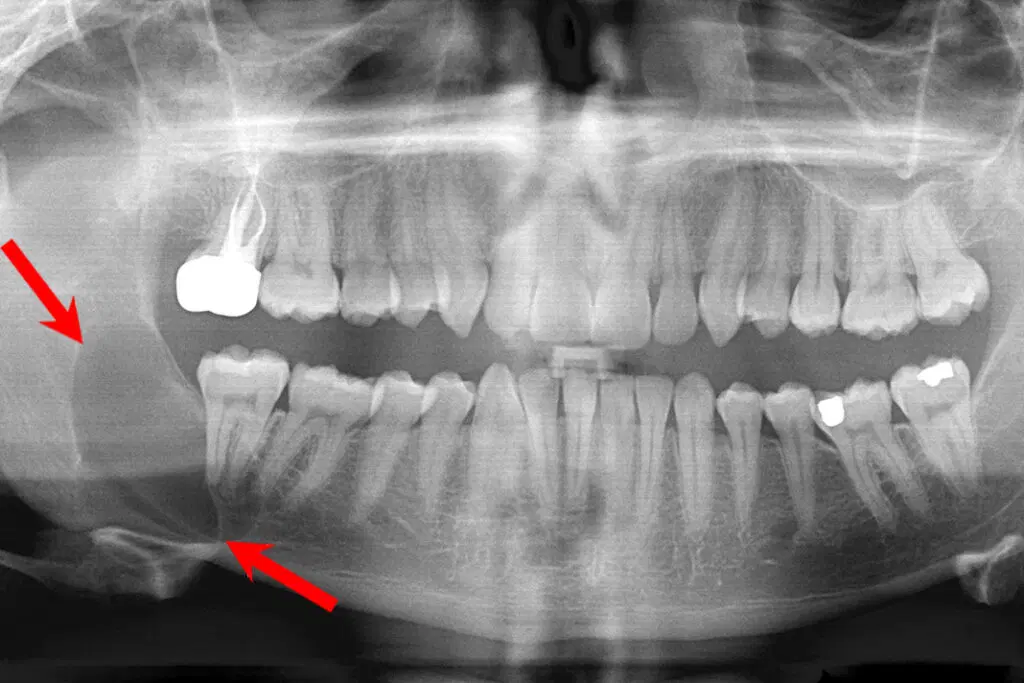
Imaging plays a key role in diagnosis. Dentists rely on panoramic X-rays and 3D CT scans. These tools reveal the size and depth of the lesion. They also show its proximity to nerves, sinuses, or roots. Some cysts lie close to the mandibular canal or maxillary sinus. Their position can complicate treatment planning. Visual data help determine whether immediate removal is necessary.
Aspiration or biopsy may be needed before deciding on a surgical approach
Not every growth behaves the same. Some contain fluid; others are solid. Aspiration helps clarify this difference. In certain cases, a biopsy confirms the lesion’s identity. These steps rule out malignancy or infection. Benign lesions may still require removal if they displace teeth or erode bone. Biopsy results guide how aggressive the surgical plan should be. The type of tissue determines the next step.
Keratocystic lesions tend to return and require wide surgical excision to prevent recurrence
Some cysts are more persistent than others. Odontogenic keratocysts, for instance, often reappear. They may invade nearby tissues without clear borders. That makes removal difficult and increases recurrence risk. Dentists often opt for more extensive surgery with these types. Margins must be clean, and follow-up is essential. Without full removal, regrowth is likely within a few years.
Dentigerous cysts commonly form around unerupted or impacted teeth, especially molars
These cysts develop in relation to tooth eruption. They often surround unerupted wisdom teeth or canines. As fluid accumulates, pressure builds in the jaw. This can damage adjacent teeth or push them out of position. If undetected, dentigerous cysts may become infected. Removal involves extracting the associated tooth and cleaning the cavity. Delayed treatment risks permanent bone damage.
Ameloblastomas are aggressive tumors that can expand without metastasizing
Not all tumors are cancerous. Ameloblastomas are locally invasive but rarely spread. They grow slowly but can disfigure the jaw if untreated. Their structure allows them to infiltrate surrounding bone. These tumors require segmental resection in severe cases. Margins must include healthy bone to prevent return. Reconstruction is often necessary, sometimes involving bone grafts or implants.
Marsupialization may be used to shrink large cysts before full surgical removal
Large cysts may not be removed in one step. Marsupialization is a technique that creates an opening in the lesion. This allows fluid to drain and pressure to drop. Over time, the cyst shrinks, making later removal easier. The process takes weeks or months and involves frequent cleaning. It’s often used when the cyst is close to nerves or major structures.
Jaw function can be preserved if removal avoids excessive bone loss
Preserving bone is a priority. Cyst or tumor removal must consider structural impact. Overly aggressive removal can weaken the jaw. This affects chewing and speech. Surgeons often use conservative methods where possible. New tools allow for minimal removal of surrounding tissue. When bone loss is inevitable, planning includes reconstruction strategies. This helps maintain quality of life after surgery.
Postoperative monitoring includes repeat imaging and symptom tracking over several months
Surgery is only one part of the process. Recovery involves follow-up imaging to confirm full removal. Some lesions recur silently. Repeat scans help detect early regrowth. Patients are advised to report numbness, swelling, or pain. Healing varies depending on lesion size and surgical technique. Scar tissue may develop, but bone often fills in slowly. Long-term observation is essential to prevent relapse.
Early diagnosis greatly improves outcome and reduces the extent of surgical intervention
Timing matters more than size. Early detection usually leads to simpler surgery. Small lesions can be removed with minimal disruption. Delayed treatment means more tissue involvement. It also raises the chance of nerve damage or tooth loss. Dentists emphasize regular imaging, especially if symptoms appear. Annual X-rays catch many issues before they progress. Prevention starts with awareness and routine care.
Source: Oral and Maxillofacial Surgeon in Dubai / Oral and Maxillofacial Surgeon in Abu Dhabi