The classification and subsequent diagnosis of Temporomandibular Joint Disorders (TMD) represent one of the most persistent and vexing challenges within the fields of dentistry, orofacial pain medicine, and neurology. Often grouped under a single, dismissive umbrella term, TMD is not a monolithic disease but a heterogeneous collection of musculoskeletal and neuromuscular conditions affecting the temporomandibular joint (TMJ), the masticatory muscles, and the associated soft tissues. The sheer diversity of presenting symptoms—ranging from an intermittent clicking sound to debilitating chronic facial pain, intractable headaches, and restricted jaw movement—means that a definitive diagnosis is rarely achieved through a single, straightforward test. Instead, the process is an investigative journey, often characterized by eliminating common mimics and integrating subjective patient reporting with objective clinical and radiographic findings. This difficulty is further compounded by the high prevalence of comorbid conditions, such such as sleep disorders, fibromyalgia, and chronic tension headaches, which frequently overlap and confound the symptom picture, making it exceedingly difficult to isolate the primary source of the patient’s distress and map a clear therapeutic path.
The Sheer Diversity of Presenting Symptoms—Ranging From an Intermittent Clicking Sound to Debilitating Chronic Facial Pain, Intractable Headaches, and Restricted Jaw Movement—Means That a Definitive Diagnosis Is Rarely Achieved Through a Single, Straightforward Test
The sheer diversity of presenting symptoms—ranging from an intermittent clicking sound to debilitating chronic facial pain
The initial and most crucial step in the diagnostic pathway for TMD is a meticulously detailed patient history. Because the condition is often cyclical and heavily influenced by psychological stressors, the clinician must move beyond a simple checklist of symptoms. The history must rigorously document the onset, duration, and frequency of the primary complaints, focusing intensely on qualitative aspects of the pain (e.g., dull ache, sharp stab, radiating, pressure) and identifying any patterns associated with function, such as chewing hard foods, speaking for extended periods, or early morning awakening. Crucially, the patient’s report must include any history of trauma (e.g., whiplash, sports injury), systemic joint disorders (e.g., rheumatoid arthritis), and any pre-existing parafunctional habits like chronic gum chewing, bruxism (tooth grinding), or clenching. A complete understanding of the patient’s lifestyle, including occupational stressors and sleep quality, often reveals the functional drivers that perpetuate the disorder, providing diagnostic clues that are missed by a purely structural examination.
The History Must Rigorously Document the Onset, Duration, and Frequency of the Primary Complaints, Focusing Intensely on Qualitative Aspects of the Pain
The history must rigorously document the onset, duration, and frequency of the primary complaints
Following the comprehensive subjective report, the clinical physical examination provides the foundational objective data. This phase is systematic, starting with palpation and auscultation. The clinician must palpate the primary muscles of mastication—the masseter, temporalis, and medial/lateral pterygoids—to identify trigger points, tenderness, and areas of hypertrophy (overdevelopment), which often indicate a myofascial pain component. The TMJ itself is auscultated (listened to, often with a stethoscope) while the patient opens and closes their jaw to identify and differentiate various joint sounds—a single ‘click’ upon opening, a reciprocal click upon closing, or the grating sound of crepitus. This assessment includes a precise measurement of the maximal incisal opening (MIO) and any deviation upon opening, as restricted movement is a key feature of both internal joint derangement and severe muscle splinting. The precise location and nature of pain elicited during these movements help distinguish between an intracapsular problem (within the joint) and an extracapsular problem (in the muscles).
The Clinician Must Palpate the Primary Muscles of Mastication—The Masseter, Temporalis, and Medial/Lateral Pterygoids—to Identify Trigger Points, Tenderness, and Areas of Hypertrophy
The clinician must palpate the primary muscles of mastication
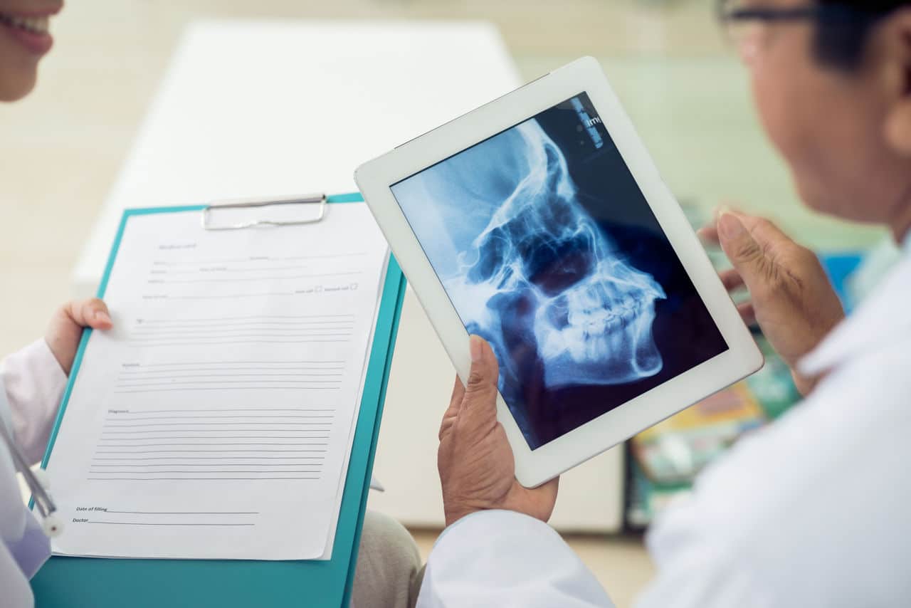
The critical step of structural differential diagnosis involves using imaging modalities to assess the integrity of the bony structures and the soft tissues of the joint. Simple panoramic or standard lateral cephalometric X-rays offer a broad overview but lack the resolution needed for fine joint detail. Computed Tomography (CT) scans are highly effective at visualizing the bony components of the joint—the mandibular condyle and the temporal fossa—allowing the clinician to identify degenerative changes, such as osteophytes (bone spurs), erosion, or flattening, which are hallmarks of osteoarthritis of the TMJ. However, CT scans are limited in their ability to visualize the joint’s crucial soft tissue structures, most notably the articular disc (meniscus). For definitive assessment of disc position, morphology, and displacement—key features of Internal Derangement—the gold standard imaging modality remains Magnetic Resonance Imaging (MRI). The MRI provides highly detailed images of the disc, ligaments, and fluid, allowing the practitioner to determine if the disc is displaced with reduction (clicking) or without reduction (locking).
The CT Scans Are Highly Effective at Visualizing the Bony Components of the Joint—The Mandibular Condyle and the Temporal Fossa—Allowing the Clinician to Identify Degenerative Changes
The CT scans are highly effective at visualizing the bony components of the joint
A significant element of the diagnostic process involves isolating the pain source through the strategic use of diagnostic local anesthetic injections. When the patient presents with a mixture of headache, ear pain, and jaw pain, it can be extremely challenging to determine which structure—the muscle, the joint capsule, or a specific nerve—is the primary generator of the pain signal. A carefully administered injection of a short-acting anesthetic directly into a suspected trigger point within the masseter or temporalis muscle can temporarily abolish the pain, confirming a myofascial etiology. Similarly, injecting anesthetic directly into the TMJ capsule can, if the pain resolves, confirm that the source is indeed intracapsular, likely due to synovitis or arthritis. This pharmacological investigation allows the clinician to move beyond observation and patient report, providing a reliable, objective physiological confirmation of the pain’s anatomical origin, which is invaluable for selecting the appropriate definitive treatment, whether it be muscle relaxants or intra-articular anti-inflammatory agents.
A Carefully Administered Injection of a Short-Acting Anesthetic Directly Into a Suspected Trigger Point Within the Masseter or Temporalis Muscle Can Temporarily Abolish the Pain, Confirming a Myofascial Etiology
A carefully administered injection of a short-acting anesthetic directly into a suspected trigger point
The diagnostic criteria used to categorize TMD have undergone significant evolution, moving toward greater standardization and reliability. The most widely accepted framework is the Diagnostic Criteria for Temporomandibular Disorders (DC/TMD). This two-axis system separates the diagnostic process into a Physical Axis (Axis I), which categorizes the primary clinical condition based on specific signs and symptoms (e.g., myalgia, disc displacement, arthralgia), and a Psychosocial Axis (Axis II), which assesses the patient’s pain-related disability, jaw function status, and presence of psychological distress (e.g., depression, somatic symptoms). The inclusion of Axis II is critical because the severity of TMD-related pain is often strongly correlated not just with the physical damage but also with the patient’s psychological coping mechanisms and central pain processing. A diagnosis should therefore be comprehensive, detailing both the structural problem and the psychosocial impact to guide a truly holistic and effective management plan.
This Two-Axis System Separates the Diagnostic Process Into a Physical Axis (Axis I), Which Categorizes the Primary Clinical Condition Based on Specific Signs and Symptoms, and a Psychosocial Axis (Axis II)
This two-axis system separates the diagnostic process into a physical axis (Axis I)
Distinguishing TMD from other causes of facial and headache pain is a necessary and highly refined step in the process, requiring the clinician to possess a broad differential diagnosis. The symptoms of TMD, particularly atypical facial pain and headaches, can closely mimic those arising from trigeminal neuralgia, dental pulpitis, otitis (ear infections), or certain cervical spine disorders. For instance, a posterior tooth with an abscess can cause referred pain that is indistinguishable from myofascial pain until a specific pulp vitality test is performed. Similarly, pain originating from the upper cervical vertebrae (C1-C3) often refers to the temporal or facial region, making a detailed physical examination of the neck and a specific cervical flexion-rotation test essential for ruling out a non-TMJ source. The final diagnosis of TMD is often achieved through a process of careful exclusion, systematically eliminating these potential mimics before concluding that the masticatory system is the true source of the discomfort.
The Symptoms of TMD, Particularly Atypical Facial Pain and Headaches, Can Closely Mimic Those Arising From Trigeminal Neuralgia, Dental Pulpitis, Otitis (Ear Infections), or Certain Cervical Spine Disorders
The symptoms of TMD, particularly atypical facial pain and headaches, can closely mimic those arising from trigeminal neuralgia
In certain instances, particularly when non-surgical treatments have failed or before surgical intervention is contemplated, arthroscopic evaluation of the joint may be employed, moving the diagnostic process from the non-invasive to the minimally invasive. TMJ arthroscopy involves inserting a small, fiber-optic camera into the joint space, allowing the surgeon to directly visualize the condition of the articular cartilage, the synovial lining, and the integrity of the disc. This direct visualization can confirm the precise nature of the internal derangement or degenerative disease with an accuracy that no non-invasive imaging technique can fully replicate. Moreover, arthroscopy often serves a dual purpose, allowing for simultaneous lavage (washing) of the joint space to remove inflammatory debris or the precise application of anti-inflammatory agents, making it an investigative tool that immediately transitions into a therapeutic one. This level of invasiveness is typically reserved for complex, refractory cases.
TMJ Arthroscopy Involves Inserting a Small, Fiber-Optic Camera Into the Joint Space, Allowing the Surgeon to Directly Visualize the Condition of the Articular Cartilage, the Synovial Lining, and the Integrity of the Disc
TMJ arthroscopy involves inserting a small, fiber-optic camera into the joint space
The use of electromyography (EMG) and other jaw movement recording devices represents a specialized layer of diagnostic assessment, primarily employed in cases where the neuromuscular component of the disorder is suspected to be dominant. EMG measures the electrical activity of the masticatory muscles at rest and during function (clinching, chewing), helping to identify muscle hypertonicity or abnormal firing patterns that contribute to chronic pain and dysfunction. For example, sustained high levels of muscle activity at rest, particularly during sleep, can confirm a diagnosis of sleep bruxism. While these instruments provide objective physiological data, they are generally not used for initial diagnosis but rather for refining the understanding of the specific functional deficit, guiding the customization of an oral appliance (splint), or objectively monitoring the success of therapeutic interventions like biofeedback or botulinum toxin injections targeting muscle hyperactivity.
EMG Measures the Electrical Activity of the Masticatory Muscles at Rest and During Function (Clinching, Chewing), Helping to Identify Muscle Hypertonicity or Abnormal Firing Patterns
EMG measures the electrical activity of the masticatory muscles at rest and during function
Ultimately, the successful diagnosis of a Temporomandibular Joint Disorder is less about finding a single positive test result and more about constructing a coherent, evidence-based narrative that connects the patient’s subjective suffering with verifiable clinical and radiographic findings. Given the high degree of psychosocial and inflammatory comorbidity, the most effective diagnostic approach is inherently multidisciplinary, often requiring input from dentists, neurologists, pain specialists, and physical therapists. This integrated, multi-modal process, utilizing everything from detailed history taking and the DC/TMD criteria to high-resolution MRI and targeted injections, is the only reliable way to sort the structural from the muscular, the primary pain from the referred pain, and the acute symptom from the chronic functional deficiency, ensuring the patient finally receives a targeted, effective treatment plan.
