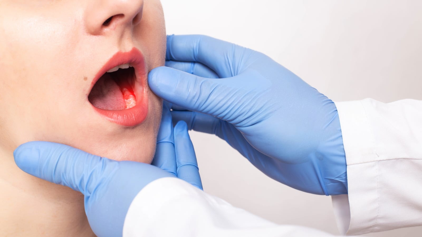
The diagnosis of oral cancer—which encompasses cancers of the lips, tongue, cheeks, floor of the mouth, hard and soft palate, and pharynx—is a life-altering event, instantly shifting the focus from routine health to complex, urgent intervention. For many patients, and indeed for the oncology team, surgery stands as the cornerstone of treatment, offering the most definitive pathway to local control and potential cure, particularly in the earlier stages of the disease. However, the prospect of an operation in such a central, critical area of the body raises a constellation of unique anxieties and complexities that extend far beyond simply removing a tumor. The oral cavity is essential for fundamental human activities—speaking, swallowing, and breathing—and is intimately linked to facial aesthetics and identity. Therefore, surgical planning in this domain becomes an exquisite balance of oncologic radicality, ensuring all malignant tissue is excised, and functional preservation, striving to maintain the patient’s quality of life following the procedure. The journey through surgical treatment is intensely personalized, dictated by the specific anatomical site of the tumor, its clinical stage, and the patient’s overall health status.
Surgery stands as the cornerstone of treatment, offering the most definitive pathway to local control and potential cure
The reality of oral cancer surgery often involves a multidisciplinary team of specialists, including head and neck surgeons, surgical oncologists, reconstructive surgeons, radiation oncologists, and specialized therapists. This collaborative approach is vital because the extent of the operation—ranging from a simple wide local excision for small lesions to complex resection involving the jawbone (mandibulectomy or maxillectomy)—will directly determine the subsequent need for adjunctive treatments and reconstructive procedures. Understanding the initial surgical plan, its anticipated margins, and the immediate post-operative implications is crucial for the patient. Unlike many other cancers, the functional and cosmetic impact of oral cancer surgery is immediate and highly visible, making pre-operative counseling and emotional preparation an essential, non-clinical part of the treatment protocol. This initial phase sets the stage for a recovery process that is often long and demanding, integrating healing with the challenge of relearning basic daily functions.
Mapping the Invasion: Pre-Surgical Diagnostic Imaging and Planning
Before the scalpel ever touches the skin, the surgical team engages in an intensive process of diagnostic imaging and strategic planning to accurately map the extent of the cancerous invasion. This is not simply about visualizing the primary tumor; it is about defining the three-dimensional boundaries of the disease and, critically, assessing the potential spread to the regional lymph nodes in the neck. Imaging modalities such as Computed Tomography (CT), Magnetic Resonance Imaging (MRI), and sometimes Positron Emission Tomography (PET) scans provide the surgeons with detailed, cross-sectional views that inform their surgical approach. The goal is to determine the optimal path for removing the tumor with adequate oncologic margins—a rim of healthy tissue surrounding the malignant lesion—to minimize the risk of recurrence.
The goal is to determine the optimal path for removing the tumor with adequate oncologic margins
The meticulous evaluation of the neck is particularly important. Oral cancers frequently metastasize (spread) to the cervical lymph nodes, often before the spread is clinically palpable. If imaging suggests involvement, or if the primary tumor is of a certain size or depth, a neck dissection will be performed concurrently with the primary tumor resection. This is a procedure to remove the lymph nodes and surrounding fatty tissue from the neck area to eliminate potential microscopic disease. The specific type of neck dissection—whether selective, modified radical, or radical—is chosen based on the pattern of expected spread, emphasizing the surgical precision required to achieve curative intent while minimizing damage to vital neck structures like nerves and blood vessels. This pre-surgical mapping is the foundation of a successful operation, ensuring that the surgeon knows precisely what tissue must be removed to secure the best possible outcome.
The Excision Phase: Understanding the Resection Margins
The core objective of the surgical operation is the complete excision of the primary tumor, a procedure known as resection. This phase is executed with strict adherence to the principle of obtaining clear margins. A clear margin means that the pathologist, after examining the excised tissue under a microscope, confirms that no cancer cells are present at the edges of the removed specimen. Achieving clear margins is the single most important predictor of local control and long-term survival in oral cancer surgery. Surgeons often rely on intraoperative frozen section analysis, where small tissue samples from the margins are rapidly analyzed while the patient is still on the operating table.
Achieving clear margins is the single most important predictor of local control and long-term survival in oral cancer surgery
If the frozen section analysis reveals cancer cells at the edge (a positive margin), the surgeon will return to the site and remove more tissue until a clear margin is established. This meticulous, time-consuming process ensures maximum safety but can occasionally lead to a larger final defect than initially planned. The actual surgical technique varies widely based on the location. A cancer of the tongue, for example, might require a partial glossectomy, while a tumor of the jawbone necessitates a mandibulectomy. These procedures are often named to reflect the anatomical structures being partially or wholly removed, highlighting the fact that oral cancer surgery is frequently ablative, meaning it involves the creation of a physical deficit that immediately demands consideration for restoration.
The Complexity of Reconstructive Surgery and Functional Restoration
The immediate aftermath of a significant cancer resection is the creation of a surgical defect, which, if left unaddressed, would severely compromise the patient’s ability to swallow, speak, and maintain facial structure. Therefore, the second, equally crucial phase of the operation is reconstruction. The sophistication of modern head and neck surgery means that the ablative and reconstructive teams often work simultaneously, or sequentially, to close the defect using the most appropriate method. Simple, small defects may be closed directly or with local tissue flaps (e.g., adjacent tongue or cheek tissue).
The sophistication of modern head and neck surgery means that the ablative and reconstructive teams often work simultaneously
However, large or complex defects—particularly those involving the jawbone, extensive soft palate, or full thickness cheek—typically require microvascular free flap reconstruction. This technique involves harvesting tissue (skin, muscle, and often bone) from a distant site on the patient’s body, such as the forearm (radial forearm flap) or the leg (fibular flap), and transplanting it to the oral cavity. The surgeon then meticulously re-connects the tiny blood vessels (arteries and veins) of the harvested tissue to blood vessels in the neck using a microscope. The fibular free flap, which includes a segment of bone, is particularly important for reconstructing the jawbone, as it provides both structural support and the potential for future dental implants, aiming to restore both form and function as closely as possible to the pre-cancer state. This phase is critical for determining the patient’s long-term functional success.
Anticipating Post-Operative Challenges: Pain and Swelling
Recovery from oral cancer surgery is invariably marked by significant post-operative challenges, primarily intense pain and considerable swelling. Given the dense nerve supply of the oral cavity and neck, pain management is a top priority and often requires a combination of narcotics, non-steroidal anti-inflammatory drugs (NSAIDs), and nerve blocks. However, relying too heavily on pain medication can interfere with early mobilization and swallowing exercises, presenting a management tightrope. The swelling is a natural, expected response to the extensive tissue manipulation, especially after a neck dissection or free flap transfer.
The swelling is a natural, expected response to the extensive tissue manipulation
This edema, or fluid retention, can be alarming to the patient and carries the immediate, albeit rare, risk of compromising the airway, which is why patients are closely monitored in the post-anesthesia care unit and intensive care setting. Furthermore, swelling within the mouth and throat is the primary impediment to resuming oral intake and clear speech in the immediate recovery phase. Managing this requires a combination of head elevation, cold compresses, and careful monitoring of drains that are often placed in the neck to remove excess fluid. The patience required during this period, as the swelling gradually subsides, is immense, marking the initial passage from acute surgical intervention to the long-term process of functional recovery.
The Critical Role of Speech and Swallowing Rehabilitation
The long-term success of oral cancer treatment is often measured not just by survival rates, but by the patient’s ability to successfully reintegrate into normal life, which is fundamentally dependent on the restoration of speech and swallowing function. Depending on the extent of the resection—especially involving the tongue, soft palate, or pharynx—patients frequently experience significant initial difficulty with dysphagia (swallowing difficulty) and dysarthria (speech difficulty). This necessitates immediate and often prolonged involvement from a speech-language pathologist (SLP).
Patients frequently experience significant initial difficulty with dysphagia and dysarthria
The rehabilitation process begins as soon as medically possible and involves specialized exercises designed to retrain the remaining musculature and compensate for missing tissue. Swallowing therapy focuses on strengthening the necessary muscles, adapting head and neck postures, and learning specific maneuvers (like the effortful swallow) to safely propel food and liquid past the reconstructed area and into the esophagus without aspiration (food entering the windpipe). Speech therapy works on maximizing articulation through new compensatory movements. This rehabilitation is not passive; it requires intense patient commitment and is an active, demanding process of rebuilding the motor skills required for two of life’s most basic and socially critical functions.
Nutritional Support and Maintaining Body Weight During Recovery
Given the difficulties with swallowing and the high metabolic demands of healing and, potentially, subsequent radiation or chemotherapy, nutritional support becomes a major pillar of post-operative care. Many patients will be discharged from the hospital with a feeding tube—either a nasogastric tube (NG) inserted through the nose or a gastrostomy tube (G-tube) placed directly into the stomach. This measure is a temporary, non-negotiable insurance policy to ensure the patient receives adequate calories and hydration, preventing malnutrition and muscle wasting.
This measure is a temporary, non-negotiable insurance policy to ensure the patient receives adequate calories and hydration
The use of a feeding tube allows the reconstructed oral tissues time to heal fully without the mechanical stress of chewing and swallowing food. While the ultimate goal is the complete and safe return to oral feeding, the patient is often encouraged to transition slowly, practicing with small amounts of puréed or soft foods under the guidance of the SLP and a registered dietitian. The dietitian plays a crucial role in tailoring the caloric and protein intake to support wound healing and maintain body mass, which can be challenging due to taste changes (dysgeusia) and dry mouth (xerostomia), common side effects of treatment. Weight maintenance is a critical metric for both physical strength and tolerance to any required follow-up treatments.
Addressing the Psychological and Social Impact of Altered Appearance
The surgical treatment of oral cancer often results in visible changes to the face and neck, and these alterations carry a profound psychological and social impact that cannot be overlooked. Disfigurement, even if subtle, can lead to issues with self-esteem, social anxiety, and depression. The mouth and face are the primary tools for emotional expression and interaction, and any perceived change can make the patient feel vulnerable or exposed. This is compounded by the difficulties in speaking and eating in public settings.
Disfigurement, even if subtle, can lead to issues with self-esteem, social anxiety, and depression
Comprehensive care must, therefore, include access to psychosocial support, such as counseling, support groups, or psychiatric referral. It is important for the patient and their family to understand that feelings of grief over the loss of function or appearance are normal and require professional attention. The goal of reconstruction is not merely to restore anatomical form, but to facilitate the patient’s return to a life where they feel comfortable and confident in social situations. Recognizing and actively treating the emotional and social wounds alongside the physical ones is vital for a truly holistic recovery and successful reintegration into society.
Adjuvant Therapies: Radiation and Chemotherapy After Surgery
For many oral cancer patients, surgery is not the final step; it is often followed by adjuvant therapies, primarily radiation therapy and sometimes chemotherapy. The decision to use these treatments is based on the findings from the surgically excised specimen, specifically the pathology report. High-risk features, such as positive or close surgical margins (cancer cells too near the edge), the presence of cancer in multiple lymph nodes, or cancer cells breaking through the lymph node capsule (extracapsular extension), are strong indicators for post-operative radiation.
The decision to use these treatments is based on the findings from the surgically excised specimen
Adjuvant therapy is administered to eliminate any remaining microscopic cancer cells that the surgery may have missed, thereby significantly reducing the risk of local or regional recurrence. When high-risk features are present, radiation is often combined with low-dose chemotherapy (chemoradiation) to further sensitize the cancer cells to the radiation. While highly effective, these treatments come with their own set of side effects, including severe dry mouth (xerostomia), swallowing difficulty, and skin reactions, which can complicate the patient’s recovery from surgery. The integration of these treatments requires careful coordination and timing to maximize oncologic benefit while managing the cumulative toxicity.
Long-Term Surveillance and The Risk of Recurrence
Once the acute treatment phase is complete, the patient enters a crucial period of long-term surveillance. The risk of recurrence—either at the original site (local recurrence) or in the adjacent area (regional recurrence)—remains a concern, particularly in the first two to three years following treatment. Furthermore, patients with a history of oral cancer, especially those with continued exposure to risk factors like tobacco or alcohol, have a heightened risk of developing a second primary cancer elsewhere in the head and neck or respiratory tract.
Patients with a history of oral cancer… have a heightened risk of developing a second primary cancer
Therefore, a rigorous follow-up schedule is instituted, typically involving frequent visits to the surgical and oncologic teams. These appointments include meticulous physical examinations of the entire oral cavity, pharynx, and neck, sometimes supplemented by regular surveillance imaging. Patient vigilance is equally essential; they are taught to be aware of and report any persistent sore spots, non-healing ulcers, lumps, or changes in voice or swallowing. This commitment to continuous monitoring and early detection is vital, as spotting a recurrence or a second primary cancer early significantly improves the chances of successful salvage treatment and long-term survival.
