Jaw asymmetry often becomes noticeable when chewing feels uneven or one side appears visually dominant. Many people live with minor asymmetry that doesn’t cause concern. However, larger differences affect appearance, bite, and jaw function. The imbalance can stem from developmental issues, trauma, or underlying joint problems. One side of the jaw may grow faster or respond differently to childhood habits. Facial appearance changes as muscle patterns adjust to uneven bone structure. Over time, the imbalance may strain joints or teeth. Diagnosing cause is essential before exploring treatment or considering any surgical intervention.
Asymmetry can develop gradually due to habits like chewing on one side or leaning the head during sleep
Asymmetry can develop gradually due to habits like chewing on one side or leaning the head during sleep. Overusing one side can cause muscles to bulk unevenly. Posture, jaw clenching, or nighttime grinding also influence bone development. These factors apply pressure that guides how the lower jaw grows over time. For younger patients, these changes are more noticeable as bones remain adaptable. In adults, soft tissues may exaggerate appearance rather than actual bone difference. Dentists may spot these trends early using bite alignment checks or panoramic imaging. Identifying the root pattern leads to better long-term management.
Genetics play a significant role when jaw asymmetry appears alongside other facial or skeletal differences
Genetics play a significant role when jaw asymmetry appears alongside other facial or skeletal differences. Some families inherit tendencies toward uneven mandibular growth or abnormal joint formation. Conditions like hemifacial microsomia or craniofacial syndromes often include asymmetric jawlines. These may affect more than just bone, including soft tissue, muscles, or nerves. Genetic cases typically appear early and may require multidisciplinary evaluation. Pediatricians, orthodontists, and surgeons often work together on diagnosis. The genetic factor does not guarantee surgery, but it helps shape the overall treatment path. In many cases, growth tracking determines the appropriate timeline for correction.
Facial trauma that affects the jaw in childhood may cause long-term asymmetry if not treated promptly
Facial trauma that affects the jaw in childhood may cause long-term asymmetry if not treated promptly. Injuries to the condyle—where the jaw connects to the skull—often lead to restricted growth. One side may recover slowly or fuse in an altered position. If unnoticed, this causes gradual shift as the other side continues normal development. Fractures that heal unevenly or without proper alignment leave lasting effects. Early imaging and surgical repair reduce future complications significantly. Without it, surgical correction later becomes more complex. Monitoring healing patterns is key to minimizing structural changes in growing bone.
Some asymmetries come from joint dysfunctions, such as temporomandibular joint disorders that alter movement over time
Some asymmetries come from joint dysfunctions, such as temporomandibular joint disorders that alter movement over time. TMJ issues cause inflammation, restricted movement, or displacement of the joint disc. These limitations may favor one side, affecting chewing and muscle development. Over months or years, this uneven motion creates bone and soft tissue changes. The joint may shift backward, compress, or flatten in one direction. Symptoms often include popping, pain, or locked jaw moments. TMJ therapy may prevent worsening, but structural asymmetry might still remain. Diagnosing joint behavior helps separate muscular causes from true skeletal imbalance.
Surgical correction usually begins with imaging like 3D scans or cephalometric analysis to understand bone positioning
Surgical correction usually begins with imaging like 3D scans or cephalometric analysis to understand bone positioning. These tools allow surgeons to visualize differences in vertical and horizontal planes. The upper and lower jaws are measured against facial symmetry lines. Planning software simulates potential movements and predicts functional improvement. These visual tools support decisions about shifting, rotating, or advancing bone segments. Accurate planning reduces complications and improves post-surgical results. A detailed blueprint guides the operating team and ensures predictable healing. Without precise imaging, subtle imbalances can be missed or overcorrected.
Orthognathic surgery repositions the jawbone by cutting and shifting specific segments into better alignment
Orthognathic surgery repositions the jawbone by cutting and shifting specific segments into better alignment. The bone is carefully separated while preserving nerves and blood vessels. Titanium plates or screws hold the pieces in place during healing. The most common techniques are bilateral sagittal split for lower jaw and Le Fort I osteotomy for upper jaw. Some procedures adjust both jaws simultaneously if the midface is also involved. The operation takes several hours and requires general anesthesia. Bone healing continues for weeks after surgery, supported by restricted movement and a soft diet.
Braces are often required before and after surgery to guide teeth into proper occlusion with the adjusted jaw
Braces are often required before and after surgery to guide teeth into proper occlusion with the adjusted jaw. Before surgery, braces decompensate the teeth, preparing them to fit correctly afterward. After surgery, the new bite needs fine-tuning for stability and comfort. This phase may last several months, depending on complexity. Braces prevent relapse by ensuring the dental arch matches the skeletal position. Aligning teeth without adjusting bone rarely solves moderate to severe asymmetry. Orthodontic planning is done jointly with surgical mapping for best outcomes. Ignoring this step risks shifting or incomplete correction postoperatively.
Recovery from jaw surgery involves swelling, restricted chewing, and regular monitoring for nerve sensitivity or relapse
Recovery from jaw surgery involves swelling, restricted chewing, and regular monitoring for nerve sensitivity or relapse. Swelling peaks within three days and fades gradually over weeks. Cold compresses and elevation help manage early discomfort. Patients follow a liquid or soft diet to protect surgical sites. Numbness or tingling may persist around the chin or lips, often improving slowly. Nerve injury is rare but possible depending on anatomy and procedure length. Follow-up visits track healing progress and stability of jaw position. Physical therapy may help with muscle coordination once movement returns.
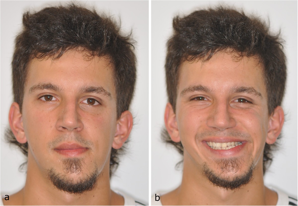
Final results become noticeable after several months as swelling subsides and bone fully integrates into the new position
Final results become noticeable after several months as swelling subsides and bone fully integrates into the new position. Patients often notice small changes daily as healing progresses. Facial balance, bite comfort, and speaking ease typically improve over time. Some need adjustments with retainers, minor contouring, or orthodontic fine-tuning. Photos taken before and after highlight how symmetry has changed. Patients regain confidence not only in appearance but also in eating and speaking. While results are stable, regular dental care remains essential to preserve alignment. Long-term satisfaction depends on collaboration, recovery habits, and maintaining follow-up appointments.